
Summary Novel methods and technology platforms developed Applications
Dynamic Contrast Enhanced Optical Imaging In silico modeling in optical molecular imaging Biomedical Spectral Imaging
Basic Principles
Spectral imaging (or chemical imaging) is an exciting new analytical advance that answers commonly asked questions such as what chemical species are in a sample, how much of each is present, and most importantly, where are they located? Through the fusion of traditional point spectroscopy with powerful microscopic and macroscopic imaging capabilities, chemical imaging spectroscopy answers all these questions simultaneously, in a single rapid measurement. Spectral imaging enables the researcher to obtain spatial and spectral information characterizing samples with unprecedented ease, speed and spatial and spectral resolution. The methodology is aimed at providing a comprehensive analysis of complex heterogeneous samples.
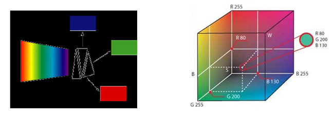
Color vision systems capture display and process, basically in real time, color images composed by three channel images, corresponding to the band-pass characteristics of the RGB primary color filters. Color images are suitable for documentation; they miss, however, significant analytical/diagnostic information since the spectral power distribution of the remitted from a target object light is sub-sampled into three, broad spectral bands. The color of a pixel can be represented as vector in a three-dimensional "color space" having the RGB values as coordinates.
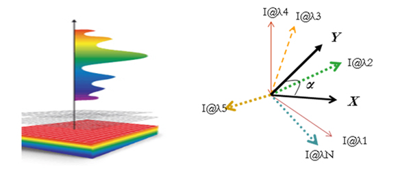
Spectroscopy finds applications in analytical chemistry since a long time. The collected spectra provide diagnostic information for the compositional status of the sample. This makes spectroscopy an indispensible tool for nondestructive analysis and for the development of novel, noninvasive diagnostic approaches. However, spectroscopy probes optical signals with high spectral resolution but with poor spatial resolution.
Spectral imaging overcomes this limitation capitalizing on its unique feature of combining the advantages of both imaging and spectroscopy (high spatial and spectral resolution) in a single instrument. Spectral Imaging systems collect a stack of images, where each image is acquired at a narrow spectral band and all together compose the so-called spectral cube. A complete spectrum can then be calculated for every image pixel, which can be otherwise represented as a vector in a "multidimensional spectral space".
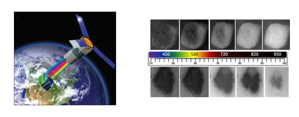
Spectral imaging camera systems can be easily adapted to a variety of optical imaging instruments such as camera lenses, telescopes, microscopes, endoscopes, etc. For this reason, applications of SI span from planetary and earth inspection (remote sensing) to internal medicine and molecular biology.
SI development and applications are increasingly migrating from the defense domain towards prevalently to civilian uses, mainly being driven by biomedical applications.
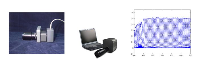
Over the past 15 years, I have developed several spectral imaging cameras, with initial intended use as research tools. In parallel and in collaboration with several scholars from a variety of application fields I had the privilege to investigate and appreciate their great potential in a variety of fields, such as microscopy, dermatology, and non -destructive analysis etc.

These cameras are competitive because they are integrated, turn-key and multimodal (all in one) solutions.
Indicative imaging modes include:
- Imaging Spectroscopy and Spectrometry
- Color Imaging and Imaging Colorimetry
- False-Color Infrared Imaging
- Multi-band Infrared Imaging
Several organizations have acquired specially tailored spectral imagers that I have designed and developed, including Johnson&Johnson, NJ, USA, Neutrogena, CA, USA Massachusetts state police (Forensic applications), Harvard University, Getty Museum in LA USA, EU Universities such as Stuttgart, Hamburg, Köln (Germany), Saragossa (Spain) etc. More Information about these systems is provided in the next paragraph.
Most recent technology developments
A. Continuously tunable, high throughput hyperspectral camera
Capitalizing on this long term past experience, we have completed the next generation of spectral cameras, based on recent innovations. The new technology is actually based on a novel continuously tunable filter/lens system (unpublished data). One major advantage is that it can now be easily integrated into mobile platforms at very low equipment cost, either as a tunable lens or as complete camera system.
Basic Specifications
Tuning step: 3nm (approx.)
FWHM: 10nm @550 nm increasing slightly with the wavelength
Spectral Range: 350-900 nm
Light throughput: 75-90%, polarization independent
Indicative applications of this new technology are:
B. The Real-Time Spectral Mapper
The Real Time Spectral Mapper, (RTSM) an advanced and unique imaging platform capable of performing the steps of capturing, processing and displaying the classification map, corresponding to millions of spectra in millisecond time regime. In other words, the output of the RTSM will be a color-coded spectral map displayed in video rate together with the image of the tissue.
The ability of displaying a diagnostic map in nearly video rates is expected to substantially advance analytical sciences and change biomedical practices. The availability of the thematic map in real time will, for example, guide medical actions, such as biopsy sampling and/or treatment, during the diagnostic scanning/interrogation of the tissue. This technology expands the applicability of spectral imaging to moving targets or to targets whose spectral characteristics are dynamically varying.
This technology has the unique feature of proving spectral acquisition, classification and mapping, @ video rates!
See “applications” for more details